IDP: Extraction of Vessels from Angiograms
Student: Titus RosuAdvisor: Prof. Dr. Rupert Lasser
Supervision by: Andreas Keil
Abstract
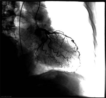
Many applications in computer-aided surgery require an automatic and exact localization of vessels in contrasted radiographs (see Fig. 1). However, the segmentation of 2D images is more difficult than segmenting 3D images because of less information contained in the data. In addition, radiographs are projections leading to occlusions (of vessels with other anatomical structures like bones as well as with other vessels). The objective of this project is the implementation and test of different methods for vessel segmentation based on eigenvalue analysis.Data
The implemented methods should be tested on different angiographic sequences (esp. rotational angiography with stationary C-arms). The images have no radial distortion since they are acquired with a flat panel detector. The pixel spacing is 0.3mm or 0.6mm which is small enough for segmenting the coronary arteries, our main target vessels.
Methods
Before segmenting vessels we improve the images by adjusting the signal-to-noise ratio and subtracting the background in the preliminary steps. The influence of the image processing [1-2] steps on the final segmentation results will be also analyzed. Most vessel segmentation techniques [4-10] are based on their linear structure and employ an eigenvalue analysis of the image intensities. Furthermore, the data will be analyzed at different scales because vessels have a varying diameter and get smaller as the degree of branching increases. Therefore, all methods have the computation of the Hessian with several derivatives of Gauss kernels on several scale steps and the subsequent eigenvalue analysis on every pixel in common. The final thresholding yields the pixels which are inside the vessels with a high probability.Objective/Evolution
Several well-known vessel segmentation/enhancement methods from the literature will be implemented and tested. The evaluation of the results should be quantitatively compared with manually segmented data. Furthermore, the accuracy improvement which is possible by specializing these algorithms to the problem at hand should be investigated.Documents
Resources and Literature
Primary:- [1] R. C. Gonzalez and R. E. Woods. Digital Image Processing. Prentice Hall, 2nd edition, 2002.
- [2] Bernd Jähne. Digital Image Processing. Springer, 5th revised and extended edition, 2002.
- [3] Tony Lindeberg. Edge detection and ridge detection with automatic scale selection. In Proc. IEEE Conf. Comp. Vis. and Pattern Recog. (CVPR), pages 465-470, June 1996.
- [4] Cemil Kirbas and Francis Quek. A review of vessel extraction techniques and algorithms. ACM Computing Surveys, 36(2):81-121, 2004.
- [5] Alejandro F. Frangi, Wiro J. Niessen, Romhild M. Hoogeveen, Theo van Walsum, and Max A. Viergever. Model-based quantitation of 3-D magnetic resonance angiographic images. IEEE Trans. Med. Imag., 18(10):946-956, 1999.
- [6] Alejandro F. Frangi, Wiro J. Niessen, Koen L. Vincken, and Max A. Viergever. Multiscale vessel enhancement filtering. In William M. Wells, III, Alan C. F. Colchester, and Scott L. Delp, editors, Proc. Int'l Conf. Med. Image Computing and Computer Assisted Intervention (MICCAI), volume 1496 of Lecture Notes in Computer Science, pages 130-137. Springer, January 1998.
- [7] Delphine Nain, Anthony Yezzi, and Greg Turk. Vessel segmentation using a shape driven flow. In Christian Barillot, David R. Haynor, and Pierre Hellier, editors, Proc. Int'l Conf. Med. Image Computing and Computer Assisted Intervention (MICCAI), volume 3216 of Lecture Notes in Computer Science, pages 51-59. Springer, 2004.
- [8] Frédéric Zana and Jean-Claude Klein. Segmentation of vessel-like patterns using mathematical morphology and curvature evaluation. IEEE Trans. Image Process., 10(7):1010-1019, 2001.
- [9] T. M. Koller, G. Gerig, Gabor Székely, and D. Dettwiler. Multiscale detection of curvilinear structures in 2-D and 3-D image data. In Proc. Int'l Conf. Comp. Vis. (ICCV), pages 864-869, June 1995.
- [10] Karl Krissian, Grégoire Malandain, Nicholas Ayache, Régis Vaillant, and Yves Trousset. Model-based detection of tubular structures in 3D images. J. Computer Vis. and Img. Understanding, 80(2):130-171, 2000.
- [11] Kostas Haris, Serafim N. Efstrtiadis, Nicos Maglaveras, Costas Pappas, John Gourassas, and George Louridas. Model-based morphological segmentation and labeling of coronary angiograms. IEEE Trans. Med. Imag., 18(10):1003-1015, 1999.
| Students.ProjectForm | |
|---|---|
| Title: | Extraction of Vessels from Angiograms |
| Abstract: | Many applications in computer-aided surgery require an automatic and exact localization of vessels in contrasted radiographs (see Fig. 1). However, the segmentation of 2D images is more difficult than segmenting 3D images because of less information contained in the data. In addition, radiographs are projections leading to occlusions (of vessels with other anatomical structures like bones as well as with other vessels). The objective of this project is the implementation and test of different methods for vessel segmentation based on eigenvalue analysis. |
| Student: | Titus Rosu |
| Director: | Prof. Dr. Rupert Lasser |
| Supervisor: | Andreas Keil |
| Type: | IDP |
| Area: | Segmentation |
| Status: | finished |
| Start: | 2007/12/13 |
| Finish: | 2008/10/14 |