|
|
in collaboration with Chirurgische Klinik und Poliklinik at Klinikum der LMU - Innenstadt and MITI Institute at Klinikum rechts der Isar
In recent years, an increasing number of liver tumor indications were treated by minimally invasive laparoscopic resection. Besides the restricted view, a major issue in laparoscopic liver resection is the precise localization of the vessels to be divided. To navigate the surgeon to these vessels, pre-operative imaging data can hardly be used due to intra-operative organ deformations caused by appliance of carbon dioxide pneumoperitoneum and respiratory motion.
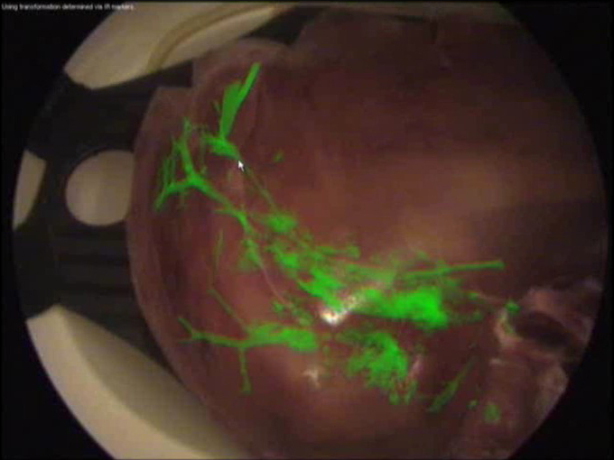
Therefore, we propose to use an optically tracked mobile C-arm providing cone-beam computed tomography imaging capability intra-operatively. After patient positioning, port placement, and carbon dioxide insufflation, the liver vessels are contrasted and a 3D volume is reconstructed during patient exhalation. Without any further need for patient registration, the volume can be directly augmented on the live laparoscope video. This augmentation provides the surgeon with essential aid in the localization of veins, arteries, and bile ducts to be divided or sealed.
Current research focuses on the intra-operative use and tracking of mobile C-arms as well as laparoscopic ultrasound, augmented visualization on the laparoscope's view, and methods to synchronize respiratory motion.
|

