Bachelor Informatics with Complementary Courses in Theoretical Medicine
Overview and Motivation
Computer science can contribute significantly to various healthcare systems. The Chair for Computer Aided Medical Procedures provides facilities to study this field within real world applications and research teams in close collaboration with medical partners at Klinikum Innenstadt and Klinikum rechts der Isar. Why is Theoretical Medicine a perfect complementary course?- Multidisciplinary lecturers: computer scientists, physicians, and physicists
- Get introduced and acquainted with an exciting, interdisciplinary field
- Learn about medical needs for computer technology
- Learn about medical processes form experts
- Learn to study requirement for real life medical applications and to model the problems adequately
- Learn to understand key medical and technical issues, and how to deal with them
- Prepare for a job in a fast-growing market
- ...
Schedule for Theretical Medicine
| Semester 3 | Anatomie I (1,5 ECTS) | Physiologie (1,5 ECTS) | Medizinische Teminologie (1,5 ECTS) | Medizinische Informatik I (1,5 ECTS) | Medizinische Statistik I (1,5 ECTS) |
| Semester 4 | Anatomie II (1,5 ECTS) | Klinische Propädeutik (1,5 ECTS) | Pathologie (1,5 ECTS) | Medizinische Informatik II (1,5 ECTS) | Medizinische Statistik II (1,5 ECTS) |
| Semester 5 | Computer Aided Medical Procedures (CAMP) I (6 ECTS) | Medizinische Signalverarbeitung (1,5 ECTS) | Medizinische Bildverarbeitung (1,5 ECTS) | ||
| Semester 6 | IDP (7ECTS) |
Recommended Courses
As optional courses for semester 5 and 6 we recommend Basic Mathematical Tools for Imaging and Visualization? (5 ECTS) and 3D Computer Vision? (6 ECTS).Exciting Thesis projects, IDPs, or Hiwi positions
There are always projects available for thesis, IDP projects and HIWI positions. One basic requirement is knowledge of C++, Matlab, and understanding of the medical context. The work will be within one of our research groups. A few examples are:
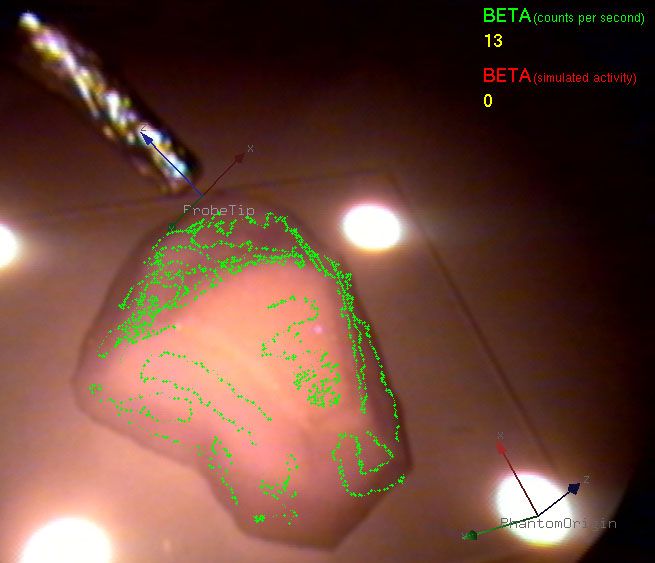
Navigated Beta Probes for Optimal Tumor Resectionin collaboration with Nuclear Medicine Department at Klinikum rechts der Isar In minimally invasive tumor resection, the goal is to perform a minimal but complete removal of cancerous cells. In the last decades interventional beta probes supported the detection of remaining tumor cells. However, scanning the patient with an intraoperative probe and applying the treatment are not done simultaneously. The main contribution of this work is to extend the one dimensional signal of a nuclear probe to a four dimensional signal including the spatial information of the distal end of the probe. This signal can be then used to guide the surgeon in the resection of residual tissue and thus increase its spatial accuracy while allowing minimal impact on the patient. |
CamC - Camera Augmented Mobile C-Armin collaboration with the Trauma Surgery Department of Klinikum Innenstadt The problem of positioning mobile C-arms, e.g. for down the beam techniques, as well as repositioning during surgical procedures currently requires time, skill and additional radiation. The Camera-Augmented Mobile C-arm (CAMC) is able to speed up the procedure, simplify its execution and reduce the necessary radiation. The C-arm is extended by a CCD camera. The CCD camera is attached to the C-arm such that it is virtually at the same location than the x-ray source. This is done by a double mirror construction. A two step calibration routine has be done only once at the moment the camera is attached. Extensions for a lightwight, but complete navigation system for pedile screw placement or implant positioning is currently under investigation. |
Deformable RegistrationObjects change their form over time. Undoing these changes makes the two images of the same object more comparable. This process of finding a transformation of one of the images, such that it gets as similar as possible to the other one is called deformable registration. Deformable registration can lead to improvement of many medical procedures, both diagnostic and interventional. The main reason is that deformations of the human body are present in many settings and for many imaging techniques. Always, when these deformations occur between different scans, there is potential for the current methods to be improved by using deformable registration techniques. Example applications include follow-up studies, image fusion from different modalities and treatment planning. The focus of our work is two-fold: we try to understand and improve the existing registration methods and to solve clinical problems by applying the developed methods. |

|
Multimodal Ultrasound RegistrationThe fusion of ultrasound data with a tomographic modality like CT or MRT is beneficial for many applications, e.g. intra-operative navigation scenarios. In particular it can also support the treatment planning and delineation of the target volume for radiation therapy. We investigate methods for image-based registration and fusion of ultrasound images with CT/MRT scans. This includes techniques for tracking, calibration and compounding for 3D freehand ultrasound, registration algorithms, as well as visualization and navigation methods. |

|
3D user interfaces for medical interventionsin collaboration with Chirurgische Klinik und Poliklinik at Klinikum der LMU - InnenstadtThis work group aims at practical user interfaces for 3D imaging data in surgery and medical interventions. The usual monitor based visualization and mouse based interaction with 3D data will not present acceptable solutions. Here we study the use of head mounted displays and advanced interaction techniques as alternative solutions. Different issues such as depth perception in augmented reality environment and optimal data representation for a smooth and efficient integration into the surgical workflow are the focus of our research activities. Furthermore appropriate ways of interaction within the surgical environment are investigated. |
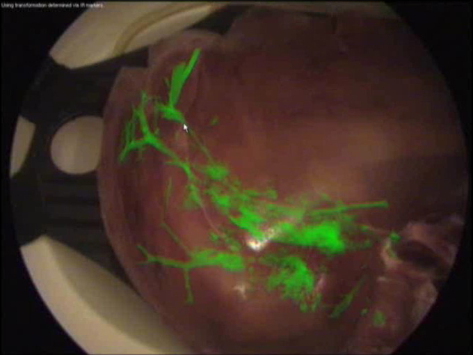
Laparoscope Augmentation for Minimally Invasive Liver Resectionin collaboration with Chirurgische Klinik und Poliklinik at Klinikum der LMU - Innenstadt and MITI Institute at Klinikum rechts der Isar In recent years, an increasing number of liver tumor indications were treated by minimally invasive laparoscopic resection. Besides the restricted view, a major issue in laparoscopic liver resection is the precise localization of the vessels to be divided. To navigate the surgeon to these vessels, pre-operative imaging data can hardly be used due to intra-operative organ deformations caused by appliance of carbon dioxide pneumoperitoneum and respiratory motion. Therefore, we propose to use an optically tracked mobile C-arm providing cone-beam computed tomography imaging capability intra-operatively. After patient positioning, port placement, and carbon dioxide insufflation, the liver vessels are contrasted and a 3D volume is reconstructed during patient exhalation. Without any further need for patient registration, the volume can be directly augmented on the live laparoscope video. This augmentation provides the surgeon with essential aid in the localization of veins, arteries, and bile ducts to be divided or sealed. Current research focuses on the intra-operative use and tracking of mobile C-arms as well as laparoscopic ultrasound, augmented visualization on the laparoscope's view, and methods to synchronize respiratory motion. |
Note: Included topic Project2D3DAngioRegHeader? does not exist yet
Contact
- Joerg Traub ( )
- Prof. Dr. Nassir Navab ( )


